When choosing an IVF clinic, most patients naturally focus on the physician, success rates, and overall reputation. However, an equally important, yet often less visible, part of that decision lies within the laboratory. Behind every successful IVF cycle is a team of embryologists and a highly controlled lab environment where eggs, sperm, and embryos are carefully handled, monitored, and protected. In many ways, a clinic is only as strong as its laboratory and the systems that support it.
This guide is designed to provide a clear, step-by-step understanding of what happens inside an IVF lab, from egg retrieval through embryo transfer. By understanding these processes, patients can ask informed questions about how their embryos are labeled, tracked, monitored, and safeguarded.
Clinics that are confident in their laboratory practices should be transparent about them. In fact, this type of information should be readily available on clinic websites, allowing patients to fully evaluate their options. If a clinic is proud of its lab, its protocols, and its technology, it should feel comfortable sharing that information openly. With this knowledge, patients are better equipped to choose a clinic not only based on outcomes, but on the quality, safety, and transparency of the laboratory practices that support their journey to parenthood.
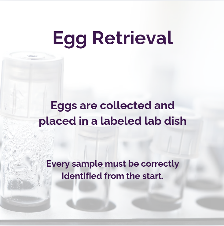
Stage 1: Egg Retrieval
Egg retrieval is the first critical step in the IVF laboratory process. After ovarian stimulation, eggs are collected using a minimally invasive ultrasound-guided procedure. The follicular fluid is immediately passed to the embryology team, where embryologists carefully search for and isolate each egg under a microscope and place it into labeled culture dishes.
The number of eggs retrieved can vary widely. In some cycles, only 2–8 eggs are collected, while in others, particularly in high responders, 20 or more eggs may be retrieved. While a higher number may seem beneficial, it also creates significant logistical and time pressures within the lab. Each egg must be identified, assessed, and prepared for fertilization within a limited time window.
Eggs cannot remain unfertilized indefinitely. After retrieval, they are typically kept in carefully controlled incubators and should ideally be fertilized within 4 to 6 hours for optimal outcomes. Beyond this window, egg quality can decline, reducing the potential for fertilization.
Eggs may be distributed across multiple culture dishes depending on quantity and treatment method. For example:
-
Mature eggs designated for ICSI are handled individually
-
Other eggs may be grouped in dishes for conventional insemination
From the moment the eggs reach the embryologist, they are working against the clock. Precision is important, but so is speed. Each decision made during this stage can directly impact fertilization success and the embryo’s future development.
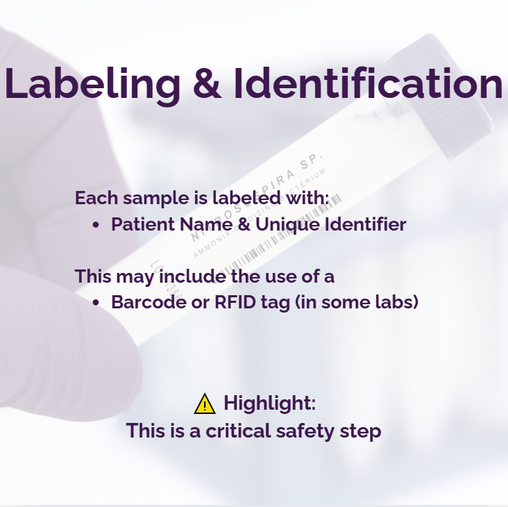
Stage 2: Labeling & Identification
Accurate labeling begins at egg retrieval and continues throughout the entire IVF process. Today, many labs are moving beyond handwritten labels to advanced digital systems.
Recent high-profile embryo mix-up cases have highlighted the risks associated with manual labeling processes. In at least one widely reported case, patient identifiers were handwritten on storage containers, and the patients had similar last names. This raises concerns about how easily human error can occur when systems rely solely on manual verification, including interpreting handwritten information. While such events are rare, they underscore the importance of strong safeguards at every step.
Solutions like eWitness by Vitrolife use barcode and RFID-enabled labels that store patient data and ensure traceability across every stage of the process.
Some clinics are also adopting biometric identification systems, such as eIVF Biometrics, which link patient identity directly to their samples, adding an additional layer of security.
These technologies:
-
Eliminate reliance on handwritten labels
-
Reduce the risk of human error
-
Create a fully auditable chain of custody
Labeling should no longer be a manual task. Increasingly, the safest environments are those where identification is digitally reinforced, continuously verified, and supported by both technology and trained professionals.
Patients are encouraged to ask their IVF clinic about the systems in place to ensure accurate identification at every stage. Clinics should clearly describe the processes and technologies used in their labs so patients can carefully evaluate their options. This is essential information and should be openly communicated, including on clinic websites.
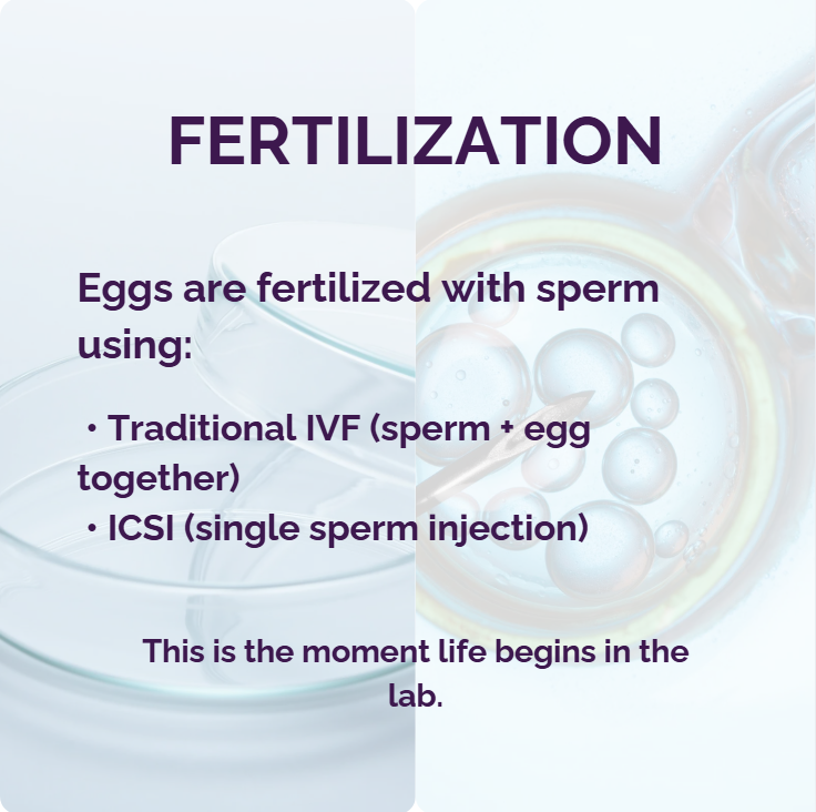
Stage 3: Fertilization
If Intracytoplasmic Sperm Injection (ICSI) has been planned, embryologists must work efficiently but carefully. Each egg must first be assessed for maturity, as only mature eggs (at the metaphase II stage) are suitable for ICSI. This means embryologists examine each egg individually before determining whether it can be injected.
At the same time, the sperm sample must also be prepared. If the sperm were previously frozen, they must be carefully thawed under controlled conditions. Embryologists must verify that the correct sample is being used and then process the sperm to isolate the healthiest, most motile sperm. This preparation step removes debris and non-viable sperm, ensuring that only the best candidates are selected for fertilization.
ICSI is a highly delicate and time-intensive procedure. An experienced embryologist may inject approximately 8 to 15 eggs per hour, depending on complexity and lab conditions. When large numbers of eggs are retrieved, labs must carefully allocate staff and timing to ensure all viable eggs are fertilized within the optimal window.
For conventional IVF (non-ICSI), eggs are placed into culture dishes and exposed to prepared sperm. Contrary to common perception, sperm is not simply “poured” over the eggs. Instead, a carefully measured concentration of processed sperm is added to the dish under controlled conditions to maximize the chance of fertilization while avoiding overcrowding or damage.
In the lab, advanced micromanipulation tools allow embryologists to perform ICSI with extreme precision. High-resolution imaging systems and automated sperm selection technologies are also improving outcomes by helping identify the healthiest sperm.
This stage represents a fusion of biology and technology, where human skill is enhanced by precision tools and data-driven insights to improve fertilization success.
Stage 4: Embryo Growth
As embryos develop over 3–6 days, maintaining a stable environment is important. Traditionally, embryos were removed from incubators for observation, but this introduced stress and variability.
Modern IVF labs now use time-lapse incubators such as the EmbryoScope+ (Vitrolife), Geri® (Genea Biomedx), and Miri® systems (Esco Medical). These systems continuously photograph embryos without removing them from their controlled environment. Many include built-in cameras that capture ongoing development, and newer artificial intelligence platforms, such as iDAScore (Vitrolife), Life Whisperer, AIVF, and Alife (Embryo Assist), analyze growth patterns, identify subtle developmental markers, and assist in ranking embryos based on their likelihood of implantation. Together, these technologies help reduce subjectivity, improve consistency, and support embryologists in managing increasingly complex IVF cycles.
This allows embryologists to:
-
Monitor development continuously
-
Identify subtle growth patterns
-
Select embryos with higher implantation potential
At the same time, this stage places significant demands on embryologists. They may be monitoring embryos from multiple patients across several days, carefully evaluating development, documenting progress, and making critical decisions. While this process is more manageable with smaller batches, cycles with 10 or more embryos per patient can substantially increase the time required for assessment and documentation.
In addition to evaluating embryo quality, embryologists must ensure that every sample removed from and returned to the incubator is correctly identified and tracked. Each movement requires careful witnessing and verification, adding another layer of responsibility during an already time-intensive process.
This shift from periodic observation to continuous AI-supported monitoring has reportedly improved both embryo selection and lab consistency.
However, it is important to note that the benefits of these technologies depend on whether they are actually implemented. Not all clinics use time-lapse imaging or AI-supported systems, and outcomes can vary based on the tools and protocols in place. Patients may wish to ask their clinic about the technologies used in the lab and how embryo development is monitored.
PGT Testing (A Parallel Step)
At the blastocyst stage (typically day 5 or 6), some embryos may be selected for preimplantation genetic testing (PGT). This involves carefully removing a small number of cells from the outer layer of the embryo (the trophectoderm), which will later form the placenta. The biopsy is performed using highly specialized equipment and requires precision to avoid damaging the embryo.
Once biopsied, the embryo is typically frozen while the sampled cells are sent to a specialized genetics laboratory for analysis. Testing may include screening for chromosomal abnormalities (PGT-A), specific inherited conditions (PGT-M), or structural rearrangements (PGT-SR).
Not all embryos are biopsied. This depends on the patient’s history, medical indications, personal preferences, and cost considerations. While PGT can provide valuable information, it is not a guarantee of pregnancy or a completely risk-free process. Patients should discuss the benefits and limitations with their clinic to determine whether this step is appropriate for their situation.
💡 Did You Know?
Not all embryos will continue to develop. It is common for a portion of fertilized eggs to stop growing before reaching the blastocyst stage. In many cases, only 30–50% of fertilized embryos make it to day 5 or 6, depending on factors such as egg quality, sperm quality, and patient age.
Stage 5: Monitoring (with Technology)
In traditional IVF labs, cryostorage monitoring relied heavily on manual processes. Staff would routinely check liquid nitrogen levels by inserting a measuring stick into each tank to assess volume, then manually top up the tanks as needed. While effective when performed consistently, this approach depends on human vigilance, timing, and accurate record-keeping. Any delay, oversight, or equipment failure between checks can pose a risk.
As an additional safeguard, most clinics maintain a reserve or emergency cryotank. In the event of a suspected tank failure or abnormal readings, embryos, eggs, or sperm can be quickly transferred to this backup tank. While this provides an important layer of protection, it still relies on timely detection and rapid response.
The importance of reliable monitoring became widely recognized following two high-profile incidents in 2018, when separate fertility clinics in the United States experienced catastrophic tank failures that resulted in the loss of thousands of eggs and embryos. In both cases, storage tanks malfunctioned, and the loss was not detected in time to prevent damage. These events led to lawsuits, increased regulatory scrutiny, and a renewed focus on improving monitoring systems and safeguards across IVF labs.
IVF labs now have access to real-time monitoring systems designed to reduce reliance on manual checks and provide continuous oversight.
These systems may include:
-
Continuous temperature sensors that track conditions inside or around the tank
-
Automated liquid nitrogen level monitoring, replacing manual dipping methods
-
Weight-based monitoring systems, which detect nitrogen loss by measuring changes in tank weight, triggering very early alerts, often before temperature changes occur
-
Remote alert systems that notify staff immediately if conditions fall outside safe ranges
Some platforms integrate all of these features into centralized dashboards, allowing staff to monitor multiple tanks across the lab 24/7.
Examples of modern monitoring technologies include:
-
ReproTech’s CryoSentinel® – real-time tank monitoring and alert system
-
TMRW Life Sciences – automated cryostorage and digital tracking platform
-
DeTech’s CryoCapture® – an integrated platform combining monitoring, witnessing, and data tracking, utilizing weight-based tank technology to detect subtle changes in liquid nitrogen levels in real time
These systems can:
-
Send instant alerts to phones or computers
-
Detect issues before temperature changes occur
-
Provide detailed audit logs for compliance and traceability
With new technology, monitoring no longer needs to be reactive; it is predictive and proactive. By combining continuous data tracking with real-time alerts, modern systems significantly reduce the risk of unnoticed failures and help protect one of the most high-risk stages of the IVF process.
Stage 6: Retrieval & Transfer
When embryos are retrieved for transfer, accuracy and identification are paramount. In clinics that do not use advanced digital systems, this process relies on manual double witnessing. At each step, from removal from storage to thawing to preparation for transfer, two embryologists must independently verify that the correct embryo is being handled. This includes checking patient names, dates of birth, and identifiers on labels and documentation.
While double witnessing is an established and important safeguard, it still depends on human attention, interpretation, and consistency. In a busy lab environment, where multiple procedures may be happening simultaneously, even highly trained professionals are working under time pressure.
Modern technology has significantly strengthened this process. Barcode and RFID-based systems, such as RI Witness (CooperSurgical) and eWitness (Vitrolife), provide automated, real-time verification. These systems track each sample throughout the lab and require confirmation at every step. If an incorrect sample is introduced, the system immediately alerts staff, preventing potential errors.
Digital tracking platforms record every movement of the embryo, while automated cryostorage systems reduce manual handling and provide precise location tracking. Together, these tools create a continuous, auditable chain of custody that goes beyond what manual systems alone can provide. However, even high-tech processes are not infallible. One of New Zealand’s largest IVF providers, Fertility Associates, has reported that in 2025, two embryos could not be located at its Auckland facility. The clinic uses the RI Witness system, which is an advanced electronic witnessing technology designed to reduce the risk of human error.
When frozen embryos are used, the process begins with careful identification and removal from cryostorage. The embryo is then thawed using controlled protocols that gradually rehydrate the cells. After thawing, embryologists assess the embryo to confirm it has survived and is suitable for transfer.
Timing at this stage is critical. Embryo thawing is carefully coordinated with the patient’s scheduled transfer. Once thawed, embryos are typically kept in controlled culture conditions for a short period, often a few hours, to allow for recovery and final assessment. They are not left outside optimal conditions, and every step is timed to maintain stability in temperature and environment.
When ready, the embryo is placed into a transfer catheter and passed to the physician for implantation under ultrasound guidance.
Despite these safeguards, past incidents have demonstrated the importance of robust identification systems. In one widely reported case, a woman in Australia underwent IVF in 1995 and gave birth to twin girls. Decades later, DNA testing revealed that she was not related to the mother who gave birth to her. An embryo mix-up had occurred. More recently, a lawsuit filed in Florida alleged that a couple’s newborn child was not genetically related to them following an IVF procedure.
Cases like these are rare but deeply distressing for everyone involved. They serve as a reminder of the extraordinary responsibility fertility clinics carry, not only to their patients, but also to the children they help bring into the world. When errors occur, the consequences are not temporary; they can affect a child’s identity, medical history, and family relationships for a lifetime.
Careful protocols, transparency, and ongoing oversight are essential to maintaining the trust that families place in reproductive medicine. This responsibility must remain paramount, even if it means clinics perform fewer procedures to ensure that every safeguard is followed and mistakes are avoided. The creation of life demands nothing less than the highest level of care and vigilance.
This stage combines the highest levels of verification, precision, and coordination between lab and clinical teams, bringing together both human expertise and technological safeguards to protect every patient and embryo.
Stage 7: Genetic Testing During or After Pregnancy
Each month, thousands of IVF cycles take place around the world, the vast majority resulting in safe, accurate, and successful outcomes. Reported errors are extremely rare. This may reflect the extraordinary dedication and precision of embryologists and clinical teams who manage these processes every day. It may also be influenced by the fact that, historically, very few patients have chosen to confirm genetic relationships after birth.
For many families, the idea of genetic testing simply never arises. It was not common practice to consider DNA confirmation after IVF. Many parents, myself included, never questioned that the child they carried and raised was genetically theirs.
However, the small number of highly publicized cases involving embryo mix-ups has demonstrated just how devastating such errors can be for everyone involved: parents, children, and clinics alike. Because the consequences are so significant, some patients are now choosing to add an additional layer of reassurance through DNA testing during or after pregnancy.
There are several options available:
- Non-Invasive Paternity Testing (NIPT):
This technology allows for DNA confirmation during pregnancy:
- Process: a simple blood draw from the mother or surrogate (containing fetal DNA) and a cheek swab from the potential father
- Accuracy: reported at 99.9%+ by many providers
Using advanced techniques, fetal DNA circulating in the mother’s or surrogate’s bloodstream can be isolated and analyzed. The baby’s DNA is then compared with that of the intended parents. The father typically provides a DNA sample through a cheek swab. Through this process, the surrogate can be excluded as a genetic contributor, while the intended mother and father can be confirmed as the baby’s genetic parents.
Timing: as early as 7–10 weeks
- Prenatal Diagnostic Testing (CVS or Amniocentesis):
These tests directly analyze fetal genetic material and can provide definitive information about the baby’s chromosomes. In specific situations, they may also be used to confirm genetic relationships, although they are more invasive and typically reserved for medical indications.
- Post-Birth DNA Testing:
After delivery, a simple cheek swab from the baby and the intended parent(s) can confirm genetic parentage with a high degree of accuracy. This is the most straightforward and commonly used method for confirmation when desired.
Final Stage
It is important to recognize the growing demands placed on IVF laboratories. IVF cycles have increased significantly year after year. With more cycles come more egg retrievals, fertilizations, embryo assessments, freezing and thawing procedures, ICSI, and embryo biopsies. Every one of these tasks is both time-sensitive and precision-dependent.
Embryologists are highly skilled professionals who are deeply committed to their work and to the families they help create. As demand increases, clinics must ensure that appropriate staffing levels and workflows are in place to support accuracy, consistency, and patient safety.
There have been ongoing industry discussions suggesting that workload and staffing ratios can influence lab performance. While there are currently no universal standards in the United States defining how many cycles an embryologist should manage or what staffing levels are appropriate, staffing levels should be an important factor to consider when evaluating which clinic and lab to work with.
This guide has been designed to help patients better understand how an IVF laboratory operates, from egg retrieval to embryo transfer, so they can ask informed questions about the processes, safeguards, and technologies used in their clinic.
Patients are encouraged to ask their IVF provider about:
- How embryos are labeled (handwritten labels should certainly be frowned upon), tracked, and monitored
- What systems are in place to prevent errors
- What technologies are used in the lab
- How the lab ensures quality and consistency across procedures
The answers to these questions can help patients make thoughtful, informed decisions about which clinic is right for them.
As the field continues to grow, it is essential that the systems supporting it evolve as well, ensuring not only technological advancement but also transparency, appropriate staffing, and a continued focus on patient safety.
We owe a duty to the children who will come from these journeys to work only with clinics whose laboratories consistently strive for the highest standards of care, safety, and transparency.
